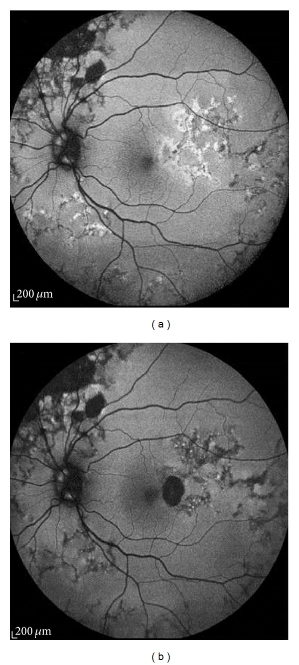
Figure 6.

Fundus autofluorescence image of left eye of a male patient with tuberculous choroiditis. (a) An ill-defined halo of hyperautofluorescence corresponding to the active lesion, giving it a diffuse, amorphous appearance. (b) Two months later, a thin rim of hypoautofluorescence appears surrounding the predominantly hyperautofluorescent lesion.
