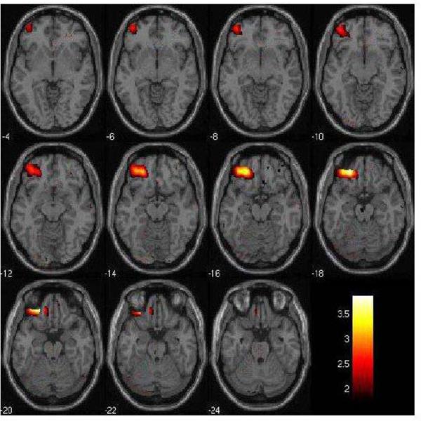
Figure 1. Left Orbitofrontal Cortex Decreases in rMDD individuals in the Fear vs. Neutral Condition.
The 2mm axial-oblique slices display the region of orbitofrontal cortex (BA11) activation decreases in the fearful vs. neutral face condition in the remitted major depressive disorder (rMDD, n=19) group compared to the healthy comparison (HC, n=20) group, at P<0.005, uncorrected. The differences survived correction for multiple comparisons (PFWE<0.05). Numbers to the left of the images are z-planes (Montreal Neurological Institute) in millimeters. The color bar shows the T values. The maxima was at coordinates x= −42mm, y= 22mm, z=−20mm; Ke= 866 voxels.

