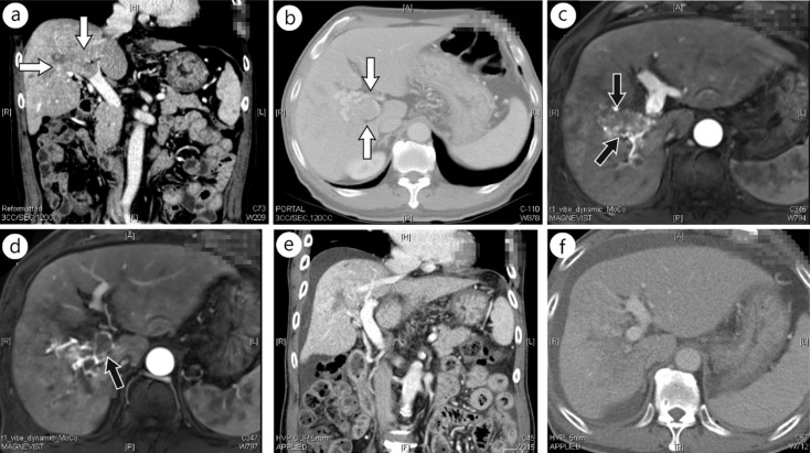
Fig. 1.
Computed tomography (CT) and magnetic resonance images (MRI) of a patient showing complete response of PVTT to concurrent chemoradiation. A 70-year-old man was diagnosed with HCC with PVTT in the right main PV trunk and its subsegmental branches. He received definitive aimed concurrent chemoradiation of 45 Gy in 25 fractions with two cycles of intra-arterial 5-fluorouracil. Pretreatment α-fetoprotein and PIVKA-II levels of 22.6 ng/ml and 11097 ng/ml decreased to 10.8 and 111 ng/ml, respectively, 3 months after chemoradiation. A nearly complete PVTT response was evident in follow-up images at 3 months. (a, b) Pretreatment CT images, (c, d) pretreatment MRIs, and (e, f) CT images 3 months after completion of chemoradiation are shown. Arrows indicate PVTT.

