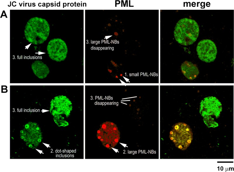
FIGURE 6.

Association of viral inclusions with PML-NBs. (A, B) Distribution of the JC virus capsid proteins VP2/VP3 (green) and the PML protein (red) in nuclei. In smaller nuclei, granular PML-NB–like structures are weakly immunoreactive to JC virus capsid proteins (A, arrow 1). As nuclei enlarge, dot-shaped viral inclusions are detected in association with large PML-NBs (B, arrow 2). In large nuclei with full viral inclusions, PML-NBs are disrupted or disappear (A, B, 3 cells with arrow 3).
