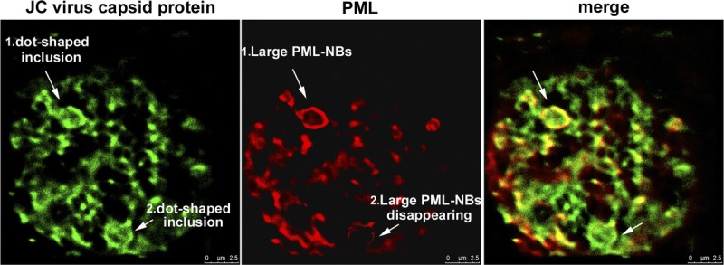
FIGURE 7.

Disruption of PML-NBs in the process of full inclusion formation. Distributions of the JC virus capsid proteins VP2/VP3 (green) and the PML protein (red) were analyzed with a superresolution GSD nanoscope. Two large PML-NBs associated with dot-shaped viral inclusions were observed in enlarged nuclei: one displayed a distinct ringlike structure more than 2 μm in diameter (arrow 1), whereas the other showed a disrupting PML-NB structure with decreased intensity of the PML protein (arrow 2).
