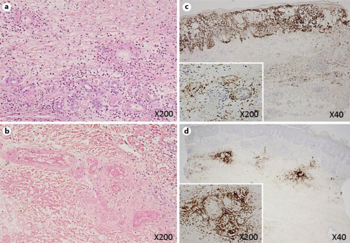
Fig. 2.
Hematoxylin- and eosin-stained section of the second biopsy specimen obtained from the bullous lesion showed leukocytoclastic vasculitis with fibrinoid degeneration in the reticular dermis and subcutaneous tissue (a). The first biopsy specimen obtained from the skin around the ulcer on the dorsum also presented perivascular infiltration of inflammatory cells and fibrinoid degeneration as well as necrotic lesions of the vessels in the dermis (b). Immunohistochemistry for VZV in the second biopsy specimen (c) and the first biopsy specimen (d) is shown. Positive staining of endothelial cells and inflammatory cells around the vessels and connective tissue in the dermis and subcutaneous tissue was observed in both specimens. On the other hand, positive staining was noted in keratinocytes only in c and was negative in d, representing the clinical features of bullous changes.

