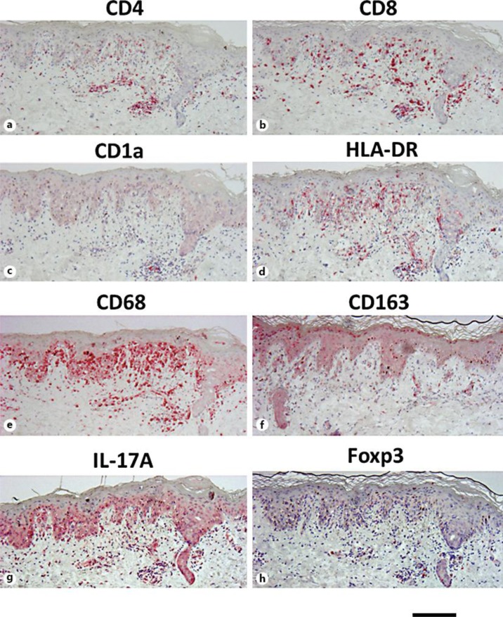
Fig. 2.
Results of immunohistochemical analysis for immunocompetent cells. Paraffin-embedded tissue samples from the patient were stained with CD4 (a), CD8 (b), CD1a (c), HLA-DR (d), CD68 (e), CD163 (f), IL-17A (g) and Foxp3 (h). CD8+ cells, instead of CD4+ cells, and HLA-DR+-activated T cells densely infiltrated into the epidermis of the erythematous lesion. CD1a+ Langerhans cells were decreased in number. While CD68+ macrophages densely infiltrated into the upper dermis, there was only slight infiltration of IL-17A+ cells and Foxp3+ cells. Bar = 250 μm.

