
Figure 5. hcASD Genes are Expressed in Midfetal Deep Layer Projection Neurons.
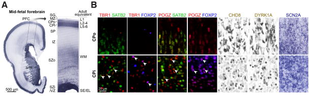
A) A coronal tissue section through the prefrontal cortex (PFC) and striatum (STR) of a midfetal forebrain was Nissl stained to visualize cells in distinct developmental zones. The darkest labeled zones are denser with cells, namely the VZ (ventricular zone) and SZi (inner subventricular zone), CPi (inner cortical plate), and CPo (outer cortical plate). The corresponding adult zones are labeled on the right of the higher magnified boxed area. B) Tissue sections of PFC areas at approximately equivalent ages (18–21 PCW) were stained with either antibodies (as labeled on top of the first six columns; fluorescence in red, green, or blue; DAB in brown) or in situ hybridization probes (last column, SCN2A). Images in the top row were taken at the boundary between CPo and MZ (marginal zone), and images in the bottom row at the boundary between CPi and SP (subplate). Arrows indicate cells colabeled for an hcASD gene and a marker gene (SATB2 or FOXP2).
SG, subpial granular zone; IZ, intermediate zone; SZo, outer subventricular zone; L, layer; WM, white matter; SE/EL, subependymal/ependymal layer.