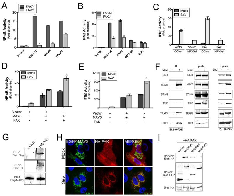
Figure 3. FAK Interacts with MAVS and other Components of RLR Signaling in Response to Virus Infection.
(A, B), Luciferase assays from FAK+/+ or FAK-/- MEFs transfected with NF-κB (A) or IFNβ (B) promoted luciferase constructs and the indicated plasmids 24 hr post-transfection. (C, D), Luciferase assays from HEK293 cells transfected with NF-κB (C) or IFNβ (D) promoted luciferase constructs and either vector, MAVS, or FAK alone or in combination. Cells were infected with SeV (25 HAU/mL) or PBS (mock) for 18 hr following transfection (24 hr) and luciferase activity measured. (E), Luciferase assay from HEK293 cells transfected with an IFNβ promoted luciferase construct and either control (CON) or MAVS siRNA. Cells were infected with SeV (25 HAU/mL) or PBS (mock) for 18 hr following transfection (48 hr) and luciferase activity measured. (F), Co-immunoprecipitation studies from HEK293 cells transfected with HA-FAK and the indicated constructs. Following transfection (48 hr), cells were infected for 18 hr with SeV (10 HAU/mL) and lysed. Lysates were subject to immunoprecipitation with antibodies directed against GFP (RIG-I, MAVS, STING, TRIF) or Flag (TRAF3, RIP1). Following washing, lysates were immunoblotted for HA-FAK (left panel) and GFP or Flag (middle panel). In parallel, lysates were immunoblotted for HA-FAK to control for the level of expression (right panel). (G), In vitro binding assays from HEK293 cells transfected with HA-FAK. Following transfection (24 hr), cells were infected for 18 hr with SeV (10 HAU/mL) and lysed. Lysates were subject to immunoprecipitation with anti-HA antibody. Following washing, recombinant Flag-MAVS was added to immunoprecipitates. Following washing, immunoprecipitates were immunoblotted for Flag-MAVS (top) and HA-FAK (middle). In parallel, lysates were immunoblotted for Flag-MAVS to control for the level of expression (bottom). (H), U2OS cells were transfected with EGFP-MAVS and HA-FAK and infected with SeV (25 HAU/mL) for 18 hr. Following infection, cells were fixed, stained for FAK (HA, red), and imaged by confocal microscopy. Blue, DAPI-stained nuclei. (I), Co-immunoprecipitation studies using HEK293 cells transfected with HA-FAK and either full-length MAVS (MAVS-FL) or N-terminal (MAVS-NT) or C-terminal (MAVS-CT) fragments of MAVS. Following transfection (48 h), cells were infected for 18 hr with SeV (10 HAU/mL) and lysed. Lysates were subject to immunoprecipitation with antibodies directed against GFP and immunoblotted for HA-FAK (top panel) or GFP (middle panel). In parallel, lysates were immunoblotted for HA-FAK to control for the level of expression (bottom panel). Data in (A -E) are shown as mean ± standard deviation. Asterisks indicate P-values of ≤ 0.05.

