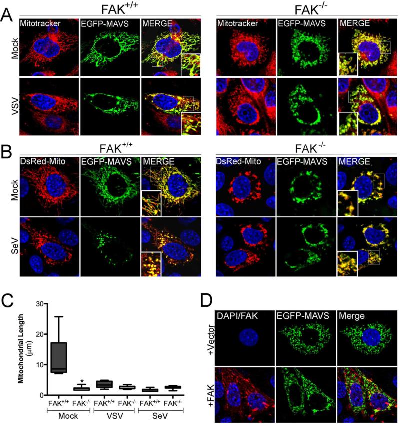
Figure 4. FAK Relocalizes to the Mitochondrial Membrane in Response to Virus Infection and Facilitates MAVS localization.
(A), FAK+/+ or FAK-/- MEFs were transfected with EGFP-MAVS and 24 hr following transfection, infected with VSV (MOI=1) for 6 hr. Following infection, cells were incubated with Mitotracker (red), fixed, and imaged by confocal microscopy. Blue, DAPI-stained nuclei. (B), FAK+/+ or FAK-/- MEFs were transfected with EGFP-MAVS and DsRed-Mito and 24 hr following transfection, infected with SeV (100 HAU/mL) for 16 hr. Following infection, cells were fixed and imaged by confocal microscopy. Blue, DAPI-stained nuclei. (C), Quantification of MAVS-associated mitochondrial length from cells shown in (A) and (B). Data are representative of a minimum of 100 individual mitochondrial filaments from at least 30 cells from three independent experiments. (D), FAK-/- MEFs were transfected with vector (top row) or wild-type FAK (bottom row) and EGFP-MAVS. Following transfection (48 hr), cells were fixed, stained for FAK (in red), and imaged by confocal microscopy. Data in (C) are shown as mean ± standard deviation. Asterisks indicate P-values of ≤ 0.05.

