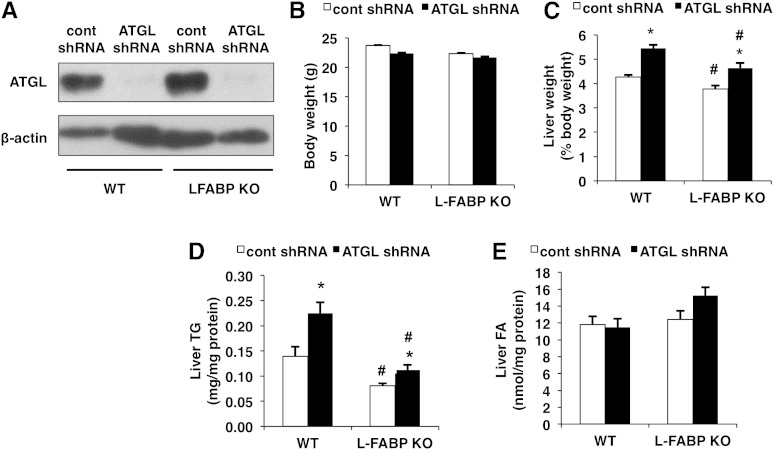
Fig. 1.
ATGL knockdown increases liver weights and TG content. WT or L-FABP KO mice, at 8–10 weeks of age, were infected with control (cont) or ATGL shRNA adenoviruses and euthanized 7 days posttransduction (n = 8–10 per group). A: Protein expression of hepatic ATGL was determined via Western blot. Body (B) and liver (C) weights, the latter expressed as a % of body weight, were measured following an overnight fast. Liver TG (D) and NEFA (E) were quantified using a colorimetric enzymatic assay. Data are presented as mean ± SEM. *P < 0.05 versus control shRNA group; #P < 0.05 versus WT mice.

