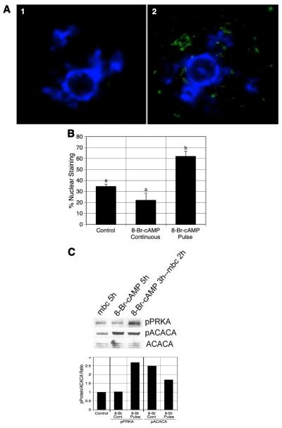
Figure 5.
Effect of 8-Br-cAMP pretreatment on PRKA activation. (A) Fluorescent nuclear staining for phospho-PRKA. mbcAMP-arrested DO were pretreated with 6 mM 8-Br-cAMP, washed free of 8-Br-cAMP, and then cultured 2 h in mbcAMP alone. GV-stage oocytes were processed for immunofluorescent staining using anti-phospho-PRKA antibody and FITC-labeled secondary antibody (green). DNA was stained with DAPI (blue). Panel 1 shows an oocyte with unstimulated PRKA, while the oocyte in panel 2 has active PRKA, indicated by punctate staining within the GV. Oocytes not exposed to primary antibody had only a faint diffuse staining throughout the oocyte and never displayed nuclear staining (not shown). (B) Quantification of germinal vesicle staining for phospho-PRKA. The frequency of nuclear (germinal vesicle) staining was compared in oocytes continuously cultured in mbcAMP or mbcAMP plus 8-br-cAMP with oocytes pulsed 3 h in mbcAMP plus 8-Br-cAMP followed by 2 h in mbcAMP alone. Groups with no common letter are significantly different. (C) Western analysis of 8-Br-cAMP-pulsed oocytes. DO treated identically to those in (B) above were processed for western analysis for pPRKA and pACACA (500 oocytes per lane). The mean pPRKA/ACACA and pACACA/ACACA ratios from 2 blots are shown, normalized to the mbcAMP alone group. Note that pPRKA increases only after an 8-Br-cAMP pulse, while pACACA levels increase after either a pulse or continuous exposure to 8-Br-cAMP. shown are the mean values for two blots.

