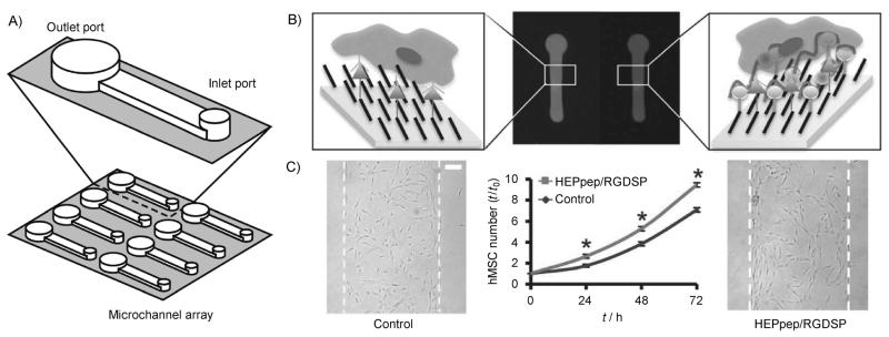
Figure 2.
Pattering SAMs using microfluidics. A) Microfluidic channels were adhered to a SAM substrate and used to locally conjugate a cell adhesion ligand (RGDSP) and a heparin-binding peptide (HEPpep). B) Schematic representation and fluorescent images of peptides patterned using microfluidics with control peptide (left) and HEPpep (right). C) hMSC number over time and bright-field photomicrographs at t= 72 h of hMSCs in regions presenting 2% RGDSP and 2% control peptide (left image), or 2% RGDSP and 2% HEPpep (right image). Scale bar=100 μm; an asterisk indicates significant difference compared to “control” at p<0.05. Adapted from ref. [6] with permission. Copyright: The Royal Society of Chemistry, 2011.

