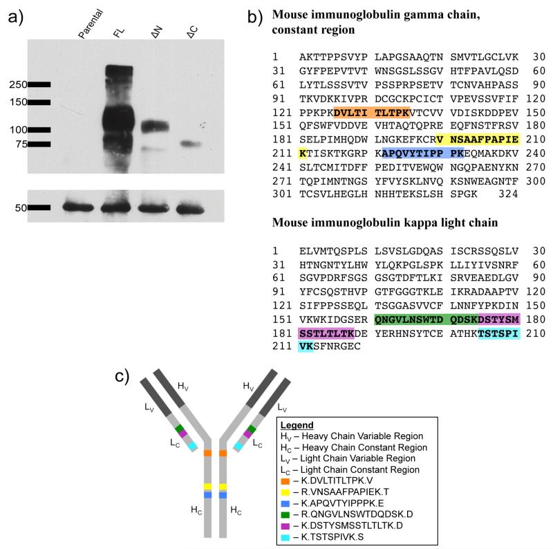
Figure 2.
Characterization of mouse IgG: (a) Western blot showing DAT (upper box) and IgGs (lower box) in the four IP samples. The blot was probed with mouse anti-HA primary antibody followed by antimouse HRP secondary antibody that recognized both the anti-HA primary antibody attached to the DAT and that which was used in the IP. (b) Peptides are highlighted within the mouse IgG constant regions. (c) Peptides are highlighted on a cartoon representation of an antibody molecule.

