Figure 6.
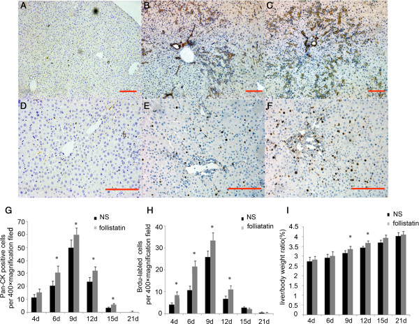
Follistatin accelerated oval cell proliferation in vivo. (A and D) negative control; Pan-CK positive hepatic progenitor cells (B and C, 100 × magnification) and BrdU positive proliferating cells (E and F, 200 × magnification) in normal sailine (NS) or follistatin treated (FST) 2-AAF/PH rats 6 day after PH were determined by immuno-histochemistry. Comparison of the number of Pan-CK positive hepatic progenitor cells (G), BrdU positive proliferating cells (H) and Liver /body weight ratio (I) in two groups of animals. Data represent mean ± SD, n = 4–6, *P < 0.05 compared to NS group. Bar = 100 um.

