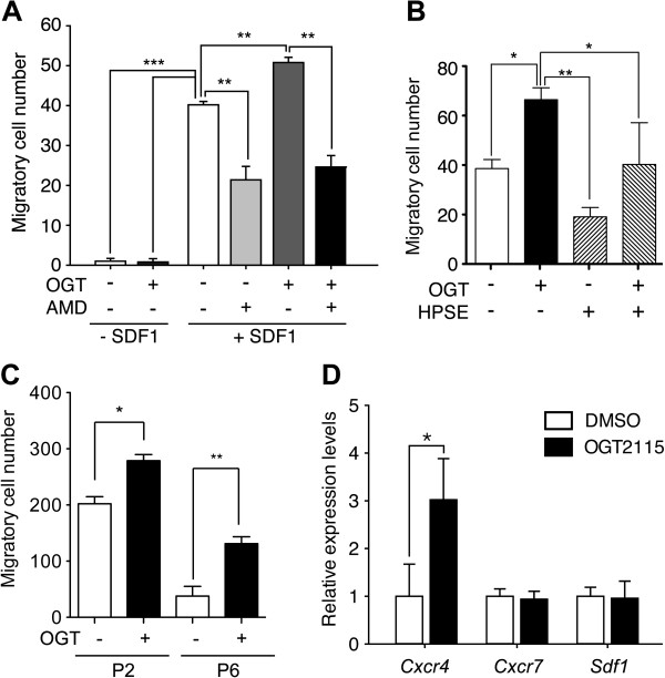
Figure 5.
The inhibition of HPSE activity potentiated the chemotaxis of BM-MSCs. (A) The chemotaxis in response to SDF-1 was assayed in P4 BM-MSCs with or without the presence of OGT2115 (OGT) and AMD3100 (AMD). The BM-MSCs were strongly mobilized by SDF-1/CXCR4 signaling axis as the migratory cells were increased with the presence of SDF-1, while the presence of OGT2115 further potentiated the migration. The addition of AMD3100, the inhibitor of SDF-1/CXCR4 signaling pathway quench the migration with or without the presence of OGT2115 (n = 3). (B) To test the specificity of the OGT2115, the transwell migration was assayed with the presence of SDF-1. The addition of mouse HPSE1 (HPSE) demonstrated a similar trend of reduction in migratory BM-MSCs as AMD3100, while the potentiation of migration by OGT2115 can be reversed by the addition of HPSE1 (n = 3). (C) The potentiation of BM-MSCS migration in response to SDF-1 by the presence of OGT2115 can also be observed in P2 and P6 BM-MSCs. (D) qPCR analysis of migration related signals indicated that HPSE inhibitor transactivated the mRNA expression level of Cxcr4 (n = 3). Error bars represent standard deviation. *P < 0.05; **P < 0.01; ***P < 0.001.

