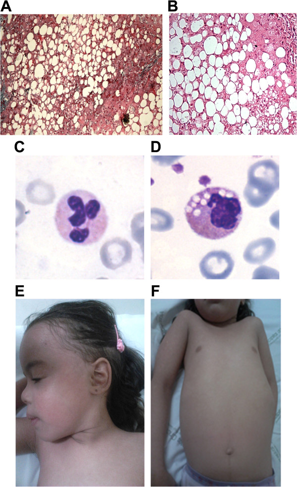
Figure 1.
Clinical phenotype of CDS patient. A and B) Liver biopsy stained with Trichrome- Magnification 100× and with Hematoxylin and Eosin (HE)-Magnification 200×; C and D) Buffy coats stained with May-Grünland Giemsa (MGG) from control subject and patient: Jordans’ bodies can be detected only in patient’s granulocytes-Magnification 100×; E and F) Erythema and fine desquamation of the skin on entire body surface.

