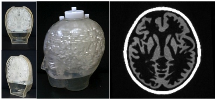
Figure 2. Left: Photographs of the Iida brain phantom used in this study.
On the left, the sections show the elaborate internal structure of the phantom with the putative ‘grey/white matter’ compartments. Right: Transaxial MPRAGE image of the brain phantom with the same slice position as shown in Fig. 7.

