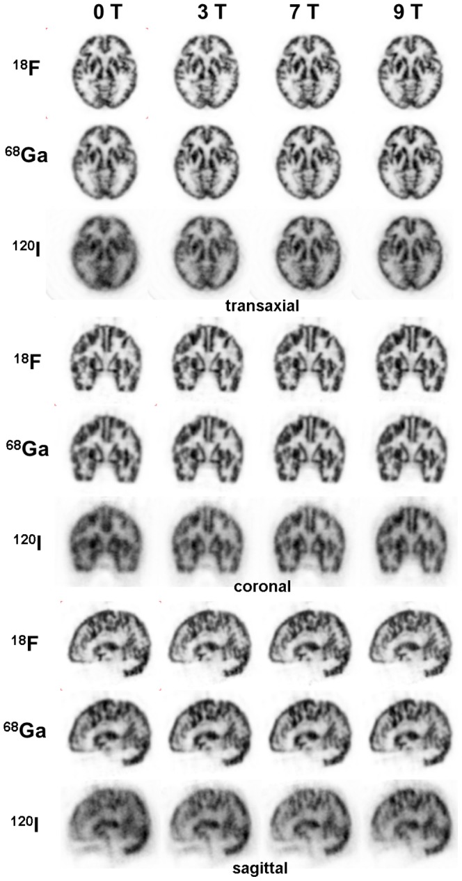
Figure 7. Transaxial, coronal and sagittal PET-images of the Iida brain phantom filled with either of three positron emitters and each measured at 0 T, 3 T, 7 T and 9.4 T.

The original reconstructed images are filtered with a 3D Gaussian filter of 3mm FWHM. Each image is scaled to its own maximum. The 18F- and 68Ga-images are scatter-corrected.
