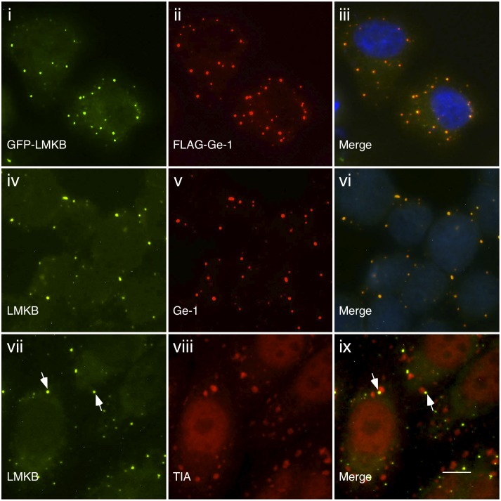
Figure 2. Indirect immunofluorescence shows that LMKB localizes to P-bodies.
GFP-LMKB (green, i) localized to discrete, dot-like structures in the cytoplasm of transfected HEp-2 cells and co-localized with co-expressed FLAG-Ge-1 (red, ii). To determine the cellular location of endogenous LMKB, rabbit anti-LMKB antiserum was used to stain Hut78 cells. LMKB (green, iv) co-localized with Ge-1 (red, v), identified using human serum 0121. After exposure to arsenite for 1 hour, TIA (a marker of stress granules) was detected in cytoplasmic granules (red, viii). LMKB (green, vii) did not co-localize with TIA in stress granules, but was instead detected in adjacent P-bodies. Merge of fluorescence in i and ii, iv and v, vii and viii is shown in iii, vi and ix. DAPI staining in iii and vi (blue) indicates the location of nuclei. White arrows in vii and ix indicate representative LMKB-containing P-bodies adjacent to stress granules. White bar in ix indicates 5.0 µm.

