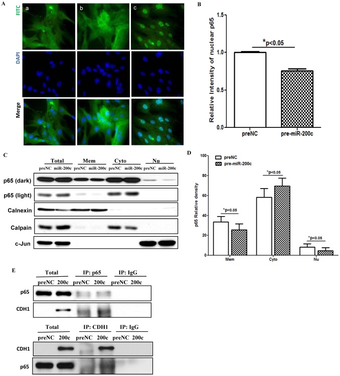
Figure 4. Gain-of function of miR-200c suppressed NF-kB signaling pathway.
Figure 4A shows immunofluorescence staining of LSMC transfected with pre-NC, (panel a), pre-miR-200c (panel b) for 48 hrs, or treated with IL-1b (5 ng/ml) for 1 h as positive control (panel c). The cells were fixed and immunostained for p65 subunit of NF-kB (green, p65) using p65 antibody and counter-stained with DAPI (blue, nucleus) with arrows indicating to NF-kB p65 nuclear immunostaining. Figure B shows mean ± SEM of relative nuclear immunostaining intensity of p65 (*p<0.05 compared to preNC). Figure 4C shows immunoblot analysis of NF-kB p65 in LSMC (3×10⁵/100 mm dish) transfected with pre-miR-200c or preNC for 48 hrs. The cells were harvested and subfractionated into membrane (Mem), cytoplasmic (Cyto), and nuclear (Nu) proteins and subjected to immunoblot analysis, with Calnexin, Calpain and c-Jun served as markers for the respective subcellular fractions. The NF-kB p65 band intensity (Fig. D) was semiquantified and reported as mean ± SEM of percentage of total p65 associated with Mem, Cyto and Nu fractions in cells transfected with pre-miR-200c or preNC (*p<0.05 when compared to preNC). Figure 4E shows immunoprecipitation and immunoblotting of LSMC total cell lysates after transfection with pre-miR-200c or preNC using p65 or CDH1 (E-cadherin) antibodies. Similar results were obtained from three sets of independent experiments.

