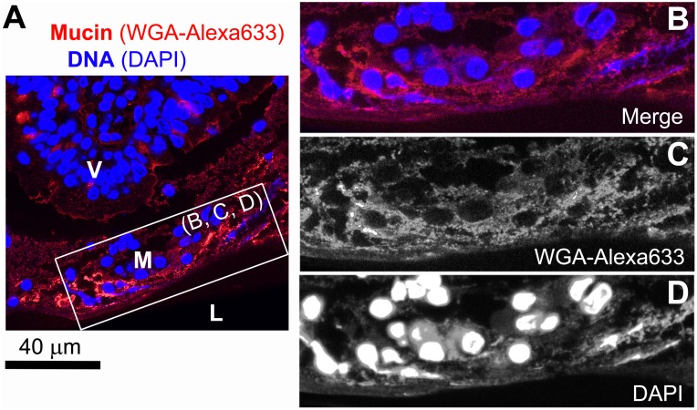
Figure 2. Thin section of pig small intestinal (jejunal) mucosa fixed with Carnoy’s solution.
(A, B) Confocal microscopy of a villus tip (V) separated from intestinal lumen (L) with a mucus layer (M). The specimen was stained for mucin with WGA-Alexa633 and with DAPI for DNA (individual channels are shown in (B) and (C), respectively).

