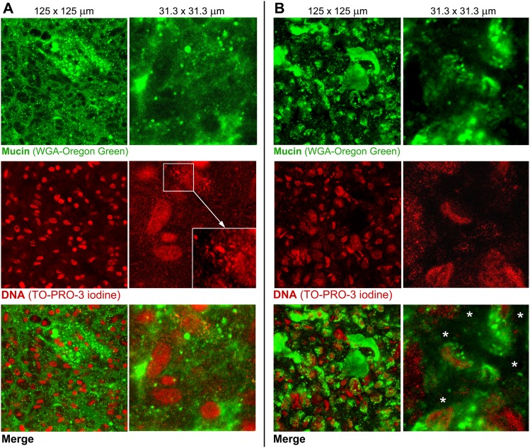
Figure 4. Comparison of the ex vivo mucus structures from pig and piglet.
Confocal microscopy images of the ex vivo small intestinal (jejunal) mucus from (A) pig and (B) piglet, acquired at two magnifications. The upper images show a green channel for mucin stained with WGA-Oregon green, the red-channel images show DNA stained with TO-PRO-3 iodine, and the bottom images are merged views of the two channels. The magnified image (A, DNA staining) highlights a progressive degradation of nuclear DNA into fine particulates. The white asterisks (B, merge) indicate the areas in the piglet mucus with apparent lower local amounts of mucin and DNA as compared to the adjacent aggregates of the two polymers.

