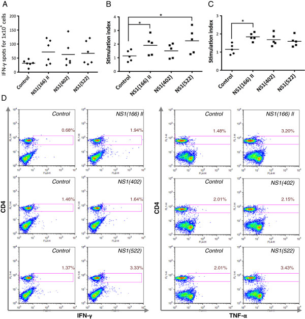
Figure 3.
NS1(166) II and NS1(522) peptides are CD4 epitopes in C57BL/6 mice. (A) No consistent significant production of IFN-γ was detected by ELISPOT to any peptide using a Mann–Whitney test. However, in individual mice significant IFN-γ production (using an unpaired Student’s t test) was detected in 3 out of 6 BTV-8-inoculated mice to peptides NS1(166) II and NS1(522) and in 2 out of 6 mice to peptide NS1(402). (B) Lymph node cells from BTV-8 inoculated mice proliferated significantly to both NS1(166) II and NS1(522) peptides. * p < 0.05 Mann–Whitney test on NS1 peptide vs control. (C) Splenocytes from BTV-8-inoculated mice consistently proliferated to the NS1(166) II peptide, whereas NS1(402) and NS1(522) peptides showed no specific proliferation. * p < 0.05 Mann–Whitney test on NS1 peptide vs control. (D) Using intracellular cytokine staining, we observed that IFN-γ and TNF-α were produced by CD4+ T cells when cultured in the presence of NS1(166) II and NS1(522) peptides, but not with NS1(402) peptide. Dot-plots are representative of at least 3 different mice. Percentages indicate the number of cytokine producing cells in the CD4+ population.

