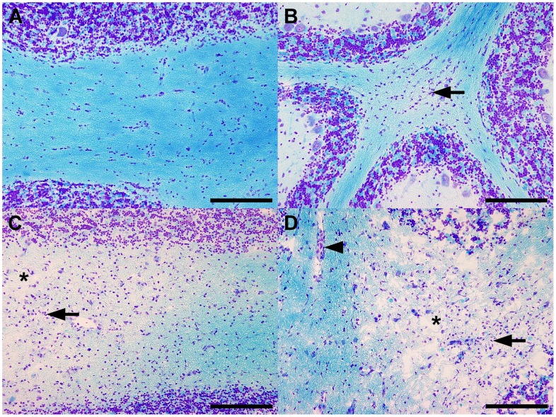
Figure 1. Pathohistological changes characteristic for the different subtypes of CDV leukoencephalitis.
(A) The cerebella of the non-infected control dogs (group 1) displayed no histological alterations. (B) The cerebella of dogs affected by acute CDV leukoencephalitis (group 2) showed focal astro- and microgliosis (arrow) and occasionally few vacuolated myelin sheaths. (C) The cerebella of dogs affected by subacute CDV leukoencephalitis with demyelination but without inflammation (group 3) exhibited focally demyelinated white matter (asterix) combined with astro- and microgliosis (arrow). (D) The cerebella of dogs affected by chronic CDV leukoencephalitis with demyelination and with inflammation (group 4) displayed focally demyelinated white matter (asterix), combined with astro- and microgliosis (arrow) as well as perivascular inflammatory infiltrates (arrowhead). Luxol fast blue-cresyl violet. Scale bars = 200 µm.

