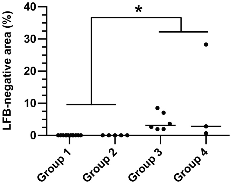
Figure 4. Multifocal demyelination is the hallmark of subacute and chronic CDV leukoencephalitis.
The dot diagram shows an increased percentage of Luxol fast blue-negative white matter area (myelin loss) in subacute CDV leukoencephalitis (group 3) and chronic CDV leukoencephalitis (group 4) as compared to controls (group 1) and acute CDV leukoencephalitis (group 2). Each point represents the percentage of Luxol fast blue-negative area relative to total white matter area within the cerebellar specimen of each individual dog. The median of each group is represented by a horizontal line. Significant differences between the groups as revealed by the Kruskal-Wallis test with independent pairwise post-hoc Mann-Whitney U-tests are marked as follows: *p≤0.05.

