
Figure 1.
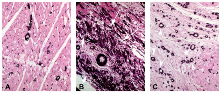
Myofibroblasts in healing myocardial infarction. A–C: Immunohistochemical staining for α-smooth muscle actin (SMA) in control canine myocardium (A) and in infarcted hearts (B – 1h coronary occlusion/7days reperfusion; C – 1h occlusion/28days reperfusion). In control hearts, α-SMA is expressed exclusively by vascular mural cells. After 7 days of reperfusion, abundant spindle-shaped α-SMA+ myofibroblasts are noted in the infarct border zone (arrows). After 28 days of reperfusion, border zone myofibroblasts are markedly reduced and α-SMA immunoreactivity is localized in vascular mural cells (arrowheads).