
Figure 7.
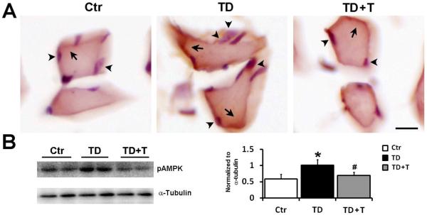
Effect of TD on AMPK phosphorylation in the peripheral skeletal muscle. Female C57BL6 mice were fed with either control or TD diet. From the 26th day on, mice in TD+T group were changed to normal liquid diet supplemented with additional benfotiamine. The mice were sacrificed and perfused on the 26th day in the TD group and the 33rd day in the TD+T and control groups, respectively. A. The expression of phosphorylated AMPKα (p-AMPKα, arrows) in skeletal muscle was demonstrated by IHC. The cells were counterstained with hematoxylin (arrowheads). Scale bar = 200 μm. B. The expression of p-AMPKα and tubulin in the skeletal muscle of the control, TD, and TD+T mice was examined by immunoblotting. The change in the expression was quantified by densitometric analysis. *p<0.05 vs. control, # p<0.05 vs. TD, n = 6 per group.