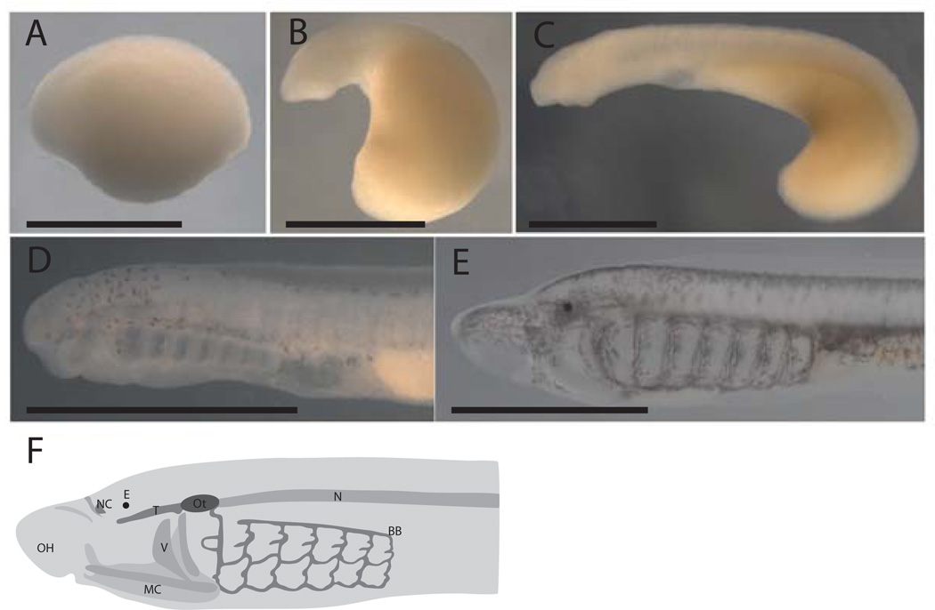
Figure 2.
External morphology during early development of the lamprey P. marinus. A. Embryo after neural rod formation, approximately Tahara Stage 20. B. Embryo at Tahara Stage 22. C. Embryo at Tahara 24.5. D. Embryo at T28 embryo. E. Proammocoete. F. Schematic of a young ammocoete, redrawn after De Beer (1937), and Langille and Hall (1988a). BB: branchial basket, E: eye, MC: mucocartilage, N: notochord, NC: nasal cartilage, OH: oral hood, Ot: Otic capsule, T: trabeculae, V: velum. Bar indicates 1 mm.

