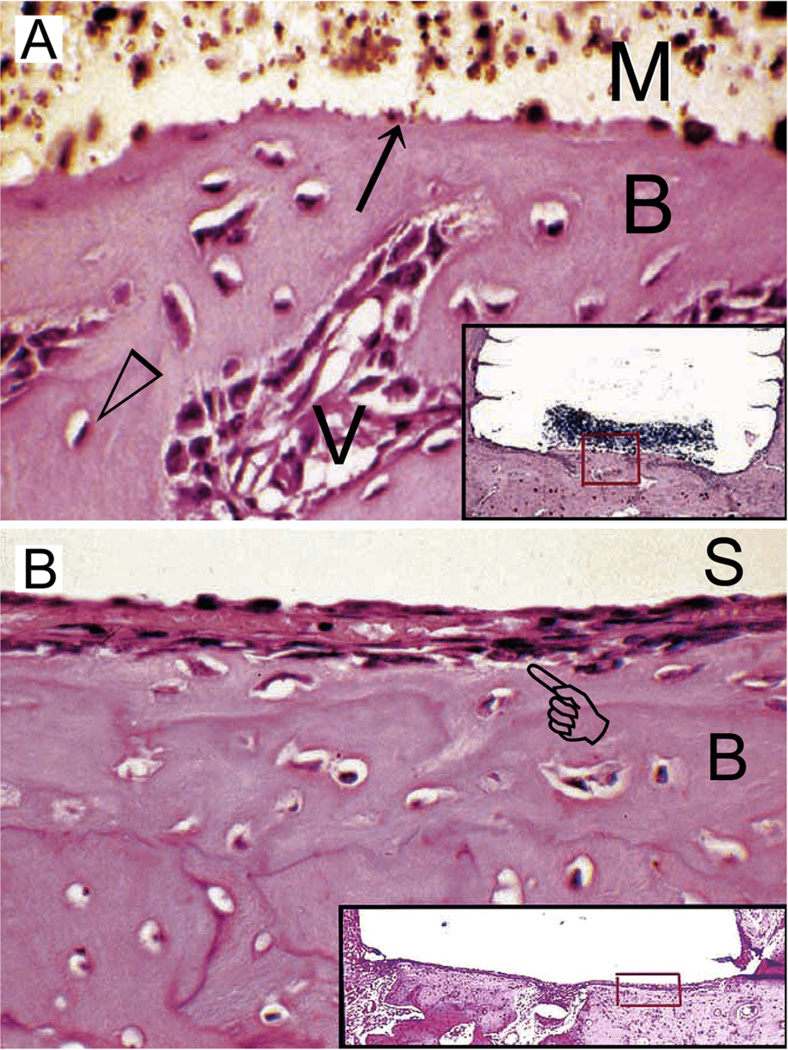
Fig. 5.
(A) Light microscopy image of the tissue response following implantation of grey MTA (ProRoot MTA) in an intraosseous site for 2 weeks. The material-hard tissue interface (arrow) shows new bone (B) apposition in direct contact with the material (M). Open arrowhead: osteocyte; V: capillary blood vessel. Inset: grey MTA-filled Teflon tube (removed, space left behind) and adjacent tissues showing remnants of grey MTA and the location (red box) from which the high magnification image is derived (haematoxylin–eosin stained section; original magnification, 250×). (B) Light microscopy image of the tissue response following implantation of grey MTA in another intraosseous site for 2 weeks. The grey MTA was lost during histological preparation (S). The bone tissue (B) has healed well, but a thin layer of fibrous connective tissue (pointer) separates the newly formed bone from direct contact with the implanted grey MTA. Inset: grey MTA-filled Teflon tube (dislodged) and adjacent tissues showing the location (red box) from which the high magnification image is derived (haematoxylin-eosin stained section; original magnification, 250×). (For interpretation of the references to colour in this figure legend, the reader is referred to the web version of this article.)
Reproduced from Saidon et al.135 with permission from the publisher; images cropped and labelled with permission from Dr. Kamran Safavi).

