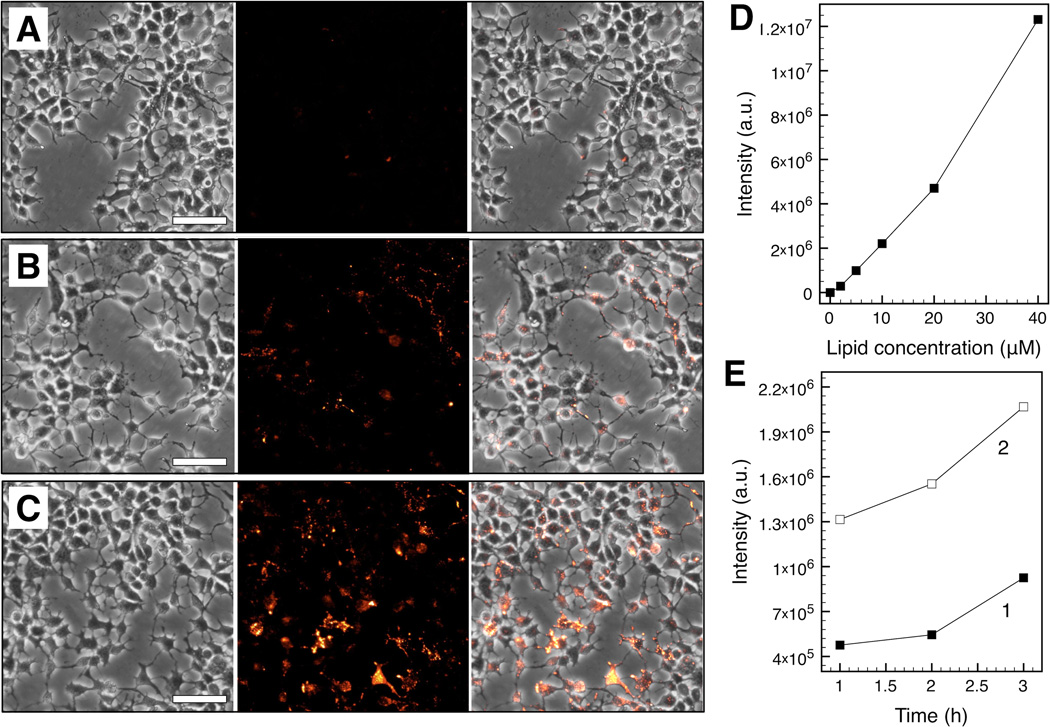
Figure 4.
In vitro dML uptake by Huh-7 cells after 24 h incubation. Corresponding bright field (left), fluorescence (middle), and merged brighfield-fluorescence (right) micrographs are shown at lipid concentrations of 5 µM (A), 20 µM (B), and 40 µM (C) (scale bar = 25 µm). Fluorescence stems from Liss Rhod PE incorporated within the dML bilayers. Total fluorescence intensity was determined from the micrographs (D) as a function of lipid concentration after 24 h incubation and (E) as a function of time at lipid concentrations of 5 µM (1) and 20 µM (2).

