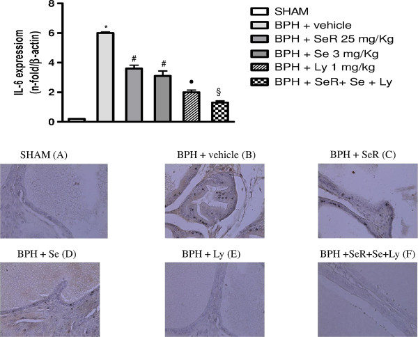
Figure 4.
IL-6 mRNA expression and Immunohistochemical analysis of PSMA.Top Panel: Expression of mRNA for IL-6. The prostate is collected from Sham rats treated with vehicle and BPH rats treated with either vehicle or SeR (25 mg/Kg) or Se (3 mg/kg) or Ly (1 mg/kg) or Ser + Se + Ly *p < 0.01 vs Sham; #p < 0.05 vs BPH + vehicle; •p < 0.01 vs BPH + vehicle; §p < 0.001 vs BPH + vehicle. Bars represent the mean ± SEM of seven experiments. Bottom panel: Immunohistochemical detection of PSMA. The prostate is collected from Sham rats treated with vehicle (A) and BPH rats treated with either vehicle (B) or SeR (25 mg/Kg) (C) or Se (3 mg/kg) (D) or Ly (1 mg/kg) (E) or SeR + Se + Ly (F). Immunohistochemistry for PSMA showed a negative reaction in sham (A) as well as in BPH treated with Ly (E) or with the combination SeR + Se + Ly (F). Differently, a moderate cytoplasmatic positivity was observed in acini and stromal cells of BPH rats treated with vehicle (B) and, in a lesser extent, of BPH rats treated with SeR (C) or Se (D). Original magnification ×400.

