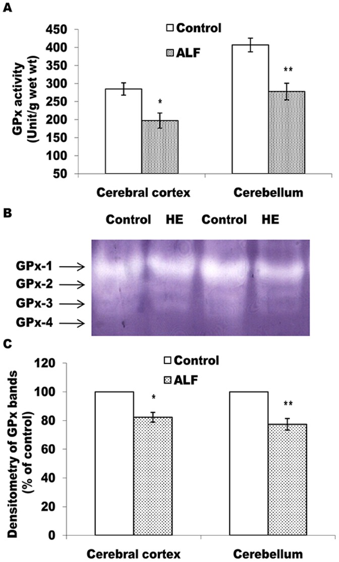
Figure 5. Level of active GPx declines in cerebral cortex and cerebellum of the ALF rats.

GPx activity measured in cell free extract (A) and non-denaturing PAGE pattern of active GPx (B and C). The technical details are same as described in Fig. 3 except 10% PAGE was used in case of B. *p<0.05; **p<0.01 (control vs ALF rats).
