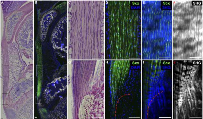
Fig. 3. Biological success criteria include 1) Scleraxis-expressing cells situated between 2) densely-packed collagen fibers aligned along the axis of tension and 3) a zonal enthesis with unmineralized and mineralized fibrocartilage.
Toluidine blue staining (A,C,G) and ScxGFP fluorescence (B,D,H) in a 4-week old murine patellar tendon depict highly aligned tendon fibroblasts within the tendon midsubstance and stacked, rounded cells within the zonal insertion site that are predominately ScxGFP+. Two photon images of tendon midsubstance (E,F) and enthesis (I,J) depict highly-aligned, densely-packed collagen fibers (second harmonic generation signal in blue and grey) within the tendon midsubstance that extend through the enthesis into the underlying bone. The tidemark (red dotted line) indicates the junction between the unmineralized and mineralized fibrocartilage. Scale bars = 100μm.

