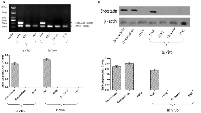
Figure 1. Expression of the exogenous endo–angio fusion gene and the activity of the fusion protein in vitro and in vivo.
(A) RT–PCR results indicated that a significant induction of endo–angio mRNA expression existed after VAE infection in vitro and in vivo. By contrast, only the internal standard control, GAPDH, could be detected in the r-HSV-, Endostar-, and PBS-treated groups. (B) Cell lysates and ECM were harvested 48 h after infection, and the orthotopically implanted tumors were harvested 10 d after injection, as described in the Materials and Methods section. The temporal pattern of the expression of endo–angio was investigated via western blot analysis of cell lysates and ECM for the in vitro experiments, and proteins extracted from the tumors for the in vivo experiments. Western blotting results indicated that a 58 kDa fusion protein recognized by the endostatin antibody was present in the VAE group, but not in the r-HSV group or the Endostar group. β-actin was detectable in all samples.

