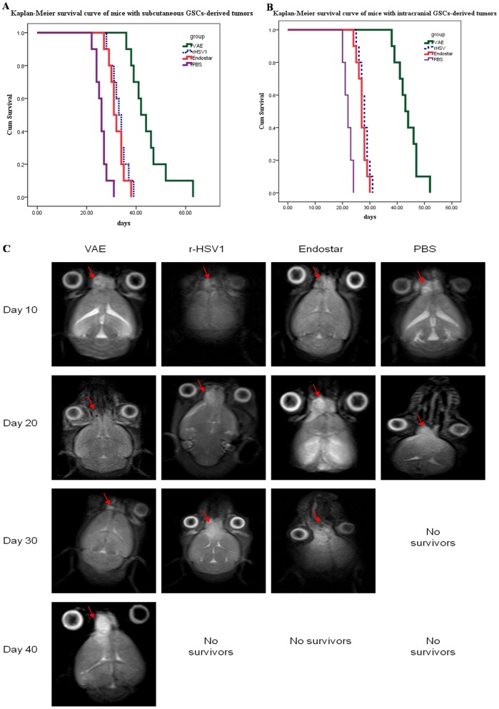
Figure 2. Increased anti-tumor efficacy of VAE compared with r-HSV, Endostar, and PBS.
(A) Kaplan–Meier survival curves of mice with subcutaneous GSC-derived tumors treated with PBS, Endostar, r-HSV, or VAE. Mice with subcutaneous GSCs-derived tumors (range = 100 mm3 to 200 mm3) were treated with PBS, Endostar, or 1×10⁷ plaque-forming units (p.f.u.) of rHSV or VAE on day 10 (n = 10/group) via direct intratumoral injection. The mice were closely monitored for tumor growth and were killed when their tumors reached 2000 mm3 or when they lost >20% of their body weight. Mice treated with VAE showed significant improvement in survival compared with mice treated with r-HSV or Endostar (P<0.001). (B) Kaplan–Meier survival curve for mice with intracranial GSC-derived tumors treated with PBS, Endostar, r-HSV, or VAE. Athymic nude mice bearing intracranial GSC-derived tumors were treated with PBS, Endostar, or 2×10⁶ p.f.u. of r-HSV or VAE 10 d after tumor cell implantation. The mice were then closely monitored for survival. Note the statistically significant improvement in median survival of mice treated with VAE compared with r-HSV- and Endostar-treated mice (P<0.001). (C) In a separate intracranial glioma experiment, mice were treated with VAE, r-HSV, Endostar, or PBS on day 10 after tumor cell implantation and examined on days 10, 20, 30, and 40 via MRI. Representative images of MRI T2-axial sections indicating the changes in tumor size (red arrows) after treatment with VAE, rHSV, Endostar, or PBS are shown. Note that all animals in the PBS-treated group developed large tumors during the treatment period. VAE-treated mice underwent complete remission, and the r-HSV- and Endostar-treated groups exhibited stable disease on day 20. After day 20, residual tumors in the VAE-treated mice regrew, and tumors in r-HSV- and Endostar-treated mice rapidly worsened (see results on day 30).

