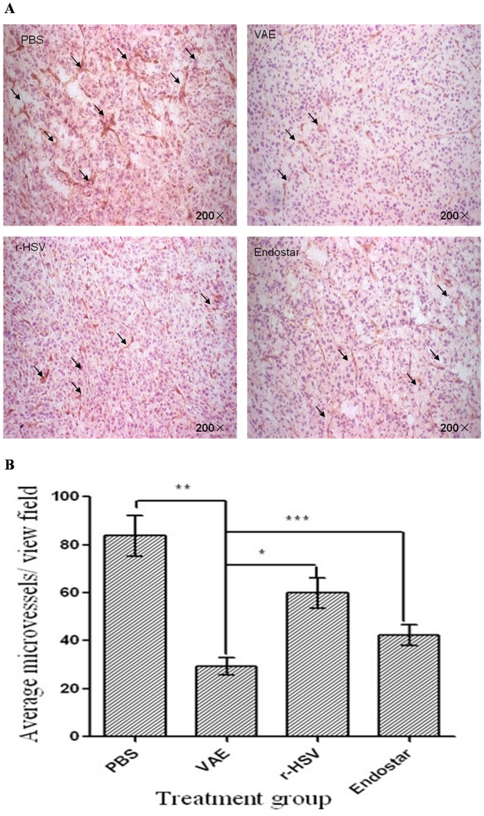
Figure 3. Reduced MVD and perfusion in animals treated with VAE compared with rHSV, Endostar, and PBS.
Mice with subcutaneous GSC-derived tumors were treated with a single dose (1×10⁷ plaque-forming units) of rHSV, VAE, PBS, or Endostar 10 d after tumor cell implantation. (A) Representative pictures of immunohistochemical staining for CD31 in tumors isolated from mice 10 d after therapy with PBS, VAE, r-HSV, or Endostar. Scale bar: 10 µm. (B) The quantification of MVD in tumors treated with PBS, VAE, r-HSV, or Endostar. Data are shown as the mean MVD ± SEM for each group (n = 2 to 4 sections/tumor and n = 10 tumors/group). Note the significant difference in MVD for VAE versus r-HSV (*P<0.001), PBS versus VAE (**P<0.001), and Endostar versus VAE (***P<0.001).

