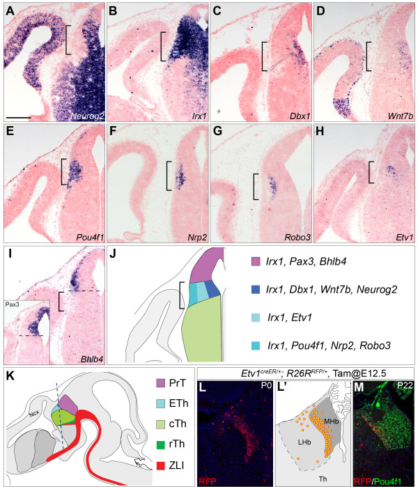
Figure 1.
Identification of molecular markers for the habenular precursor domain. (A-I) In situ hybridization (ISH) for the indicated markers on coronal sections of wildtype embryos at embryonic day (E)12.5. The inset in I shows ISH for Pax3. Brackets indicate the presumptive habenular domain; the dashed line in I demarcates the border between the pretectum and epithalamus. (J,K) Schematic representation of coronal and sagittal sections of E12.5 brains, respectively. The dashed line in K indicates the plan of sections. (L-M) Contribution of descendants (RFP+) of Etv1-expressing cells that are labeled at E12.5 to the medial habenula (MHb) in Etv1+/creER; R26R+/RFP at post-natal day (P)0 (L) or at P22 (M). The distribution of the fate-mapped cells is summarized in L’ and the MHb is marked by Pou4f1 immunoreactivity (M). cTh, caudal thalamus; ETh, epithalamus; LHb, lateral habenula; PrT, pretectum; rTh, rostral thalamus; ZLI, zona limitans intrathalamica. Scale bar in (A): 200 μm (A-I); 250 μm (L); 196 μm (M).

