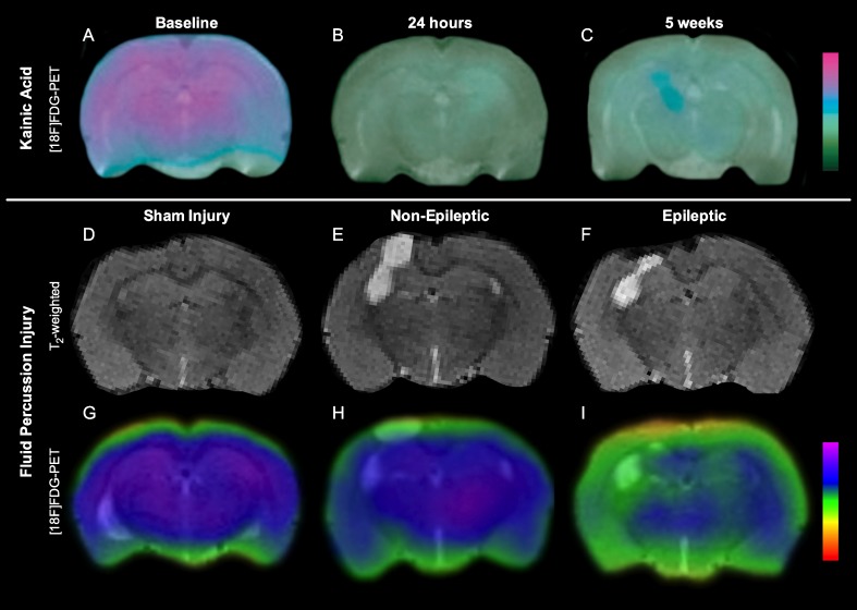
Fig. 1.
Volumetric and [18F]FDG–positron emission tomography (PET) changes in kainic acid model of temporal lobe epilepsy and fluid percussion injury model of traumatic brain injury (TBI). There is persistent hypometabolism, and progressive degeneration after kainic acid-induced status epilepticus (A–C; adapted from [29]). Although all rats suffer progressive neurodegeneration after fluid percussion injury (D–F), rats that develop post-traumatic epilepsy display persistent hypometabolism at 3 months after TBI (G–I; adapted from [11])

