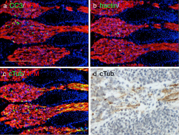
Figure 5.
TubulinΔCsp6 highlights degenerating axon tracts during normal developmental pruning. Adjacent sections from embryonic day 14.5 mice were immunolabeled with CC3 (a), fractin (b) and tubulinΔCsp6 (cTub) (c) (green) along with neurofilament (2H3; red). All three antibodies reveal a subset of apoptotic cell bodies in the ganglion, but only cTub prominently labels axons emanating from the ganglia (c,d). Notice that cTub does not label any structures in adjacent non-neural somites (above and below axon tracts). Scale = 200 μm.

