Figure 1.

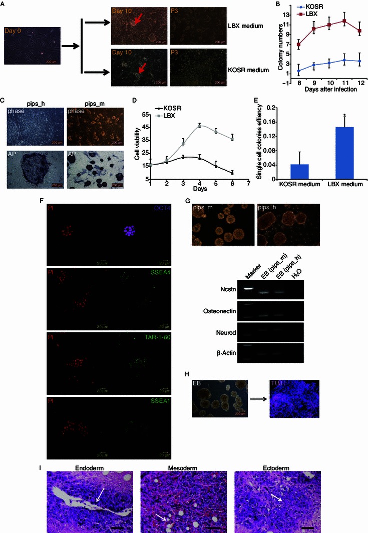
Efficient generation of mouse ESCs-like piPSCs in LBX medium. (A) Timeline for generating piPSCs in LBX and KOSR medium. Day 0 shows PEFs before infected with OSKM retrovirus, on the second day the infected PEFs were plated on MEF feeders with LBX and KOSR mediums. Day 10 shows PEFs infected 10 days later and colonies were yielded (red arrows). In LBX medium the colonies looked domed and were picked for digesting to derive cell lines. In KOSR medium the colonies looked flattened and were picked for cutting to derive cell lines. P3 shows the cell lines in KOSR and LBX mediums had been passaged for 3 times. Scale bars are 200 μm. (B) Colony number. Colonies were counted from 8 to 12 days after infection in LBX and KOSR medium, respectively. (C) Top, phase morphology of piPSCs derived in KOSR (pips_h) and LBX (pips_m) mediums. Bottom, piPSCs clones were stained with alkaline phosphatase kit. Scale bars are 200 μm. (D) Cell viability analysis for different cell lines. KOSR and LBX show the cell viability in KOSR and LBX medium. (E) Single cell colony formation. Cell lines derived in LBX medium had higher colony formation ability than that derived in KOSR medium. *P < 0.05. (F) Pips_m cells clones were stained with pluripotency markers. Positive OCT4 (purple), SSEA4 (green), TRA-1-60 (green) and SSEA1 (green) were observed. DNA was stained with propidium iodide (PI, red). Shown were examples from pips-n-3. Scale bars are 20 μm. (G) In vitro embryoid body formation. Top, pips_m and pips_h cells (from pips-n-3 and pips-5 respectively) could form EB both, scale bars are 200 μm. Bottom, endoderm (Ncstn), mesoderm (Osteonectin) and ectoderm (Neurod) markers were detected by RT-PCR. (H) Directed differentiation of pips_m cell derived EBs into neural linage. Left, EBs from examples of pips-n-3. Right, differentiated neurons were stained with Tuj1 (purple). DNA was stained with Hoechst (blue). (I) Teratoma formation of pips_m cells (from pips_m cell line pips-n-3). Tissues exhibiting all three germ layers were presented on the teratoma dissection slices identified by staining with haematoxylin and eosin. Left, endoderm with glands; Middle, mesoderm with fat tissues; Right, ectoderm with nervous tissues. Scale bars are 500 μm
