
Figure 2.
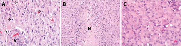
The diagnosis of glioblastoma (WHO grade IV) was rendered on histopathologic review. Histopathologic evaluation revealed a hypercellular astrocytic neoplasm which infiltrated the surrounding brain parenchyma. Mitotic activity (arrows) was abundant and microvascular proliferation (designated V) was present (A). Necrosis was encountered in the specimen, including pseudo-palisading necrosis (designated N) (B). While not a dominant appearance, focally the tumor had features of epithelioid glioblastoma (C).