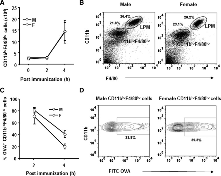
Figure 3. Antigen uptake and antigen-induced alterations in peritoneal macrophages.
Male and female SJL mice were immunized i.p. with FITC-OVA. Peritoneal cells were isolated at indicated times p.i. (A) CD11bloF4/80lo cell numbers in males and females in naïve (0 h) and at 2 and 4 h p.i. Data represent three to seven individual mice analyzed/time-point. (B) CD11b and F4/80 expression identify LPM and CD11bloF4/80lo cells. Data represent at least three experiments with similar results. (C) Percentages of FITC-OVA+ CD11bloF4/80lo cells at indicated times (n=3). (D) Percentage of OVA+ cells at 4 h p.i. Data represent at least three experiments with similar results.

