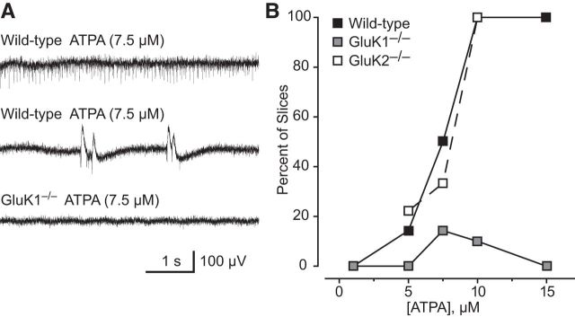
Figure 10.
Epileptiform discharges induced by ATPA in extracellular recordings in the BLA of brain slices from wild-type, GluK1−/−, and GluK1−/− mice. A, Discharges in a slice from a wild-type mouse after perfusion with 7.5 μm ATPA (top). ATPA fails to evoke such discharges in a slice from a GluK1−/− mouse (bottom). B, Percentage of slices from wild-type and GluK1−/− mice exhibiting epileptiform discharges after superfusion with various concentrations of ATPA. Each point represents 6–10 slices; 50 male 4- to 6-week-old mice were used.

