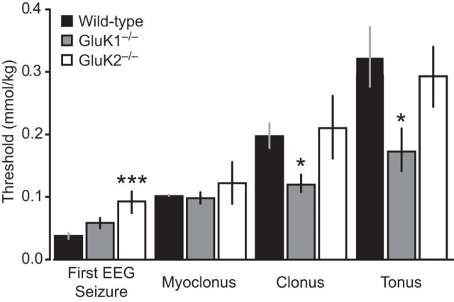
Figure 5.
Thresholds for first EEG seizure and behavioral signs during slow intravenous infusion of kainate. Kainate (25 mm) was infused at a rate of 0.03 ml/min through the lateral tail vein of wild-type, GluK1−/−, and GluK2−/− mice. Bars indicate mean ± SEM threshold for induction of the first EEG seizure, myoclonus, clonus, and tonus. Each bar represents 8–10 mice. *p < 0.05 with respect to wild-type group. ***p < 0.001 with respect to wild-type group.

