
Figure 7.
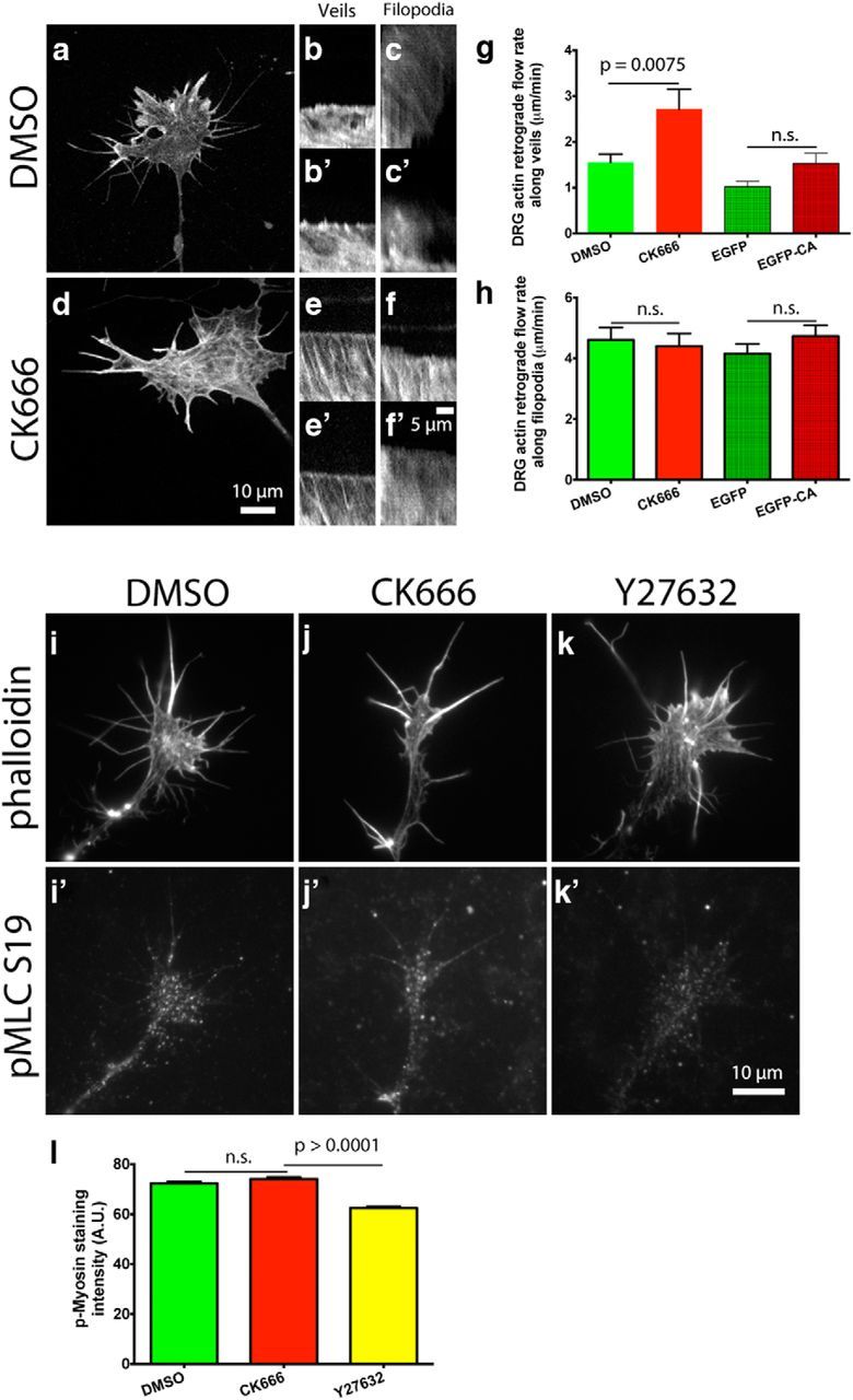
Arp2/3 inhibition increases the actin retrograde flow rate at the leading edge of growth cones on laminin in a myosin II-independent manner. a, d, E7 DRG neurons were transfected with mCherry-actin and grown overnight on laminin. Arp2/3 was inhibited by coexpression of EGFP-CA with mCherry-actin, or by 50 μm CK666 treatment 4 h before imaging. Growth cones were imaged in a spinning disc confocal and kymographs made from these time-lapse videos. b, b′,e, e′, Representative kymographs along veils and (c, c′,f, f′) filopodia of DMSO- and CK666-treated growth cones. g, h, Quantification of the actin retrograde flow rate along veils and filopodia. i–k′, As a control, DRG growth cones on L1 were treated with DMSO, 50 μm CK666, or 10 μm Y27632 for 4 h, fixed, and stained with phalloidin and an antibody against phospho myosin light chain (Ser19), (l) and anti-pMLC average fluorescent intensity quantified. Graphs show mean ± SEM.