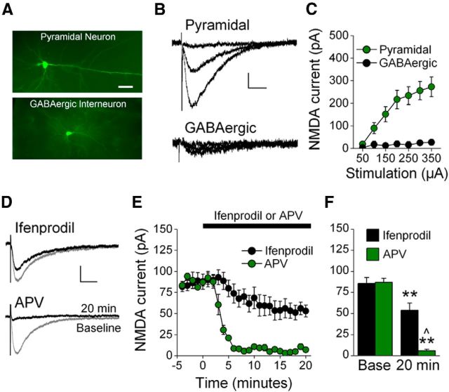
Figure 1.
Characterization of NMDAR EPSCs in IL-mPFC. A, Photomicrographs of a biocytin-filled pyramidal neuron and GABAergic interneuron. Scale bar, 100 μm. B, Example traces of NMDAR EPSCs evoked by 50, 150, and 350 μA stimulation of IL-mPFC neurons. C, NMDAR EPSCs were larger in pyramidal neurons as compared with GABAergic interneurons. D, Example traces of IL-mPFC NMDAR-dependent EPSCs before (gray) and after (black) pyramidal neurons were treated with ifenprodil (n = 7) or APV (n = 3). E, Ifenprodil significantly reduced evoked EPSCs in pyramidal neurons, and APV abolished evoked EPSCs. F, Baseline and final 5 min of recording were individually averaged; **p < 0.01 compared with baseline, ∧p < 0.01 compared with ifenprodil-treated neurons. Scale bars: vertical, 50 pA; horizontal, 50 ms. Error bars indicate SEM.

