Figure 4.

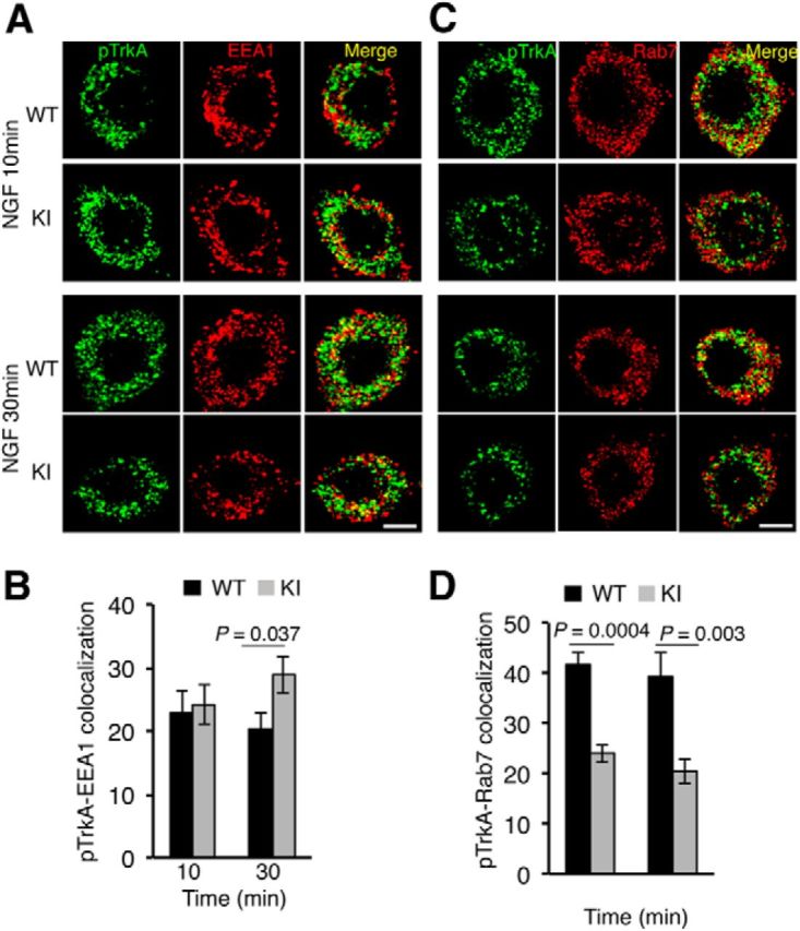
Defective trafficking of active TrkAP782S in NGF-stimulated DRG neurons. A, Colocalization of pTrkA with EEA1 compartments upon NGF treatment in WT and KI DRG neurons. Immunofluorescence was performed as described in Material and Methods. Images were taken with a confocal microscope. Scale bar, 10 μm. B, Quantification of pTrkA colocalization in EEA1 endosomes. Images were processed using ImageJ and the percentage of colocalization was quantified. Data are presented as means ± SEM; p values were calculated using a two-tailed Student's t test (n = 9–10 neurons/time point). C, Colocalization of pTrkA with Rab7 compartments upon NGF treatment in WT and KI DRG neurons was performed as described in A. Scale bar, 10 μm. D, Quantification of pTrkA colocalization in Rab7 endosomes was performed as described in B. Data are presented as means ± SEM; p values were calculated using a two-tailed Student's t test (n = 10 neurons/time point).
