Figure 7.
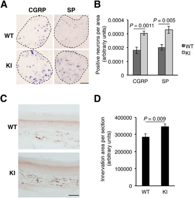
Increased CGRP and substance P (SP) expression and skin innervation in TrkAP782S mice. A, Enhanced expression of CGRP and substance P in KI mice. In situ hybridizations using antisense probes against CGRP and substance P in lumbar DRGs from P0.5 pups. Sense probes for CGRP and substance P did not show any staining. Representative pictures of DRGs from WT and KI mice. Scale bar, 100 μm. B, Quantification of positive neurons for CGRP and substance P in L3-5 DRGs from WT and KI P0.5 pups. The area of each DRG section was quantified with ImageJ software and the amount of positive neurons was counted. The number of sections quantified for CGRP was five for WT and nine for KI, and the number of sections for substance P was five for WT and six for KI. Results are presented as means ± SEM; p value was calculated using a two-tailed Student's t test. C, Skin innervation in TrkAP782S mice is increased. Skin from the hindpaws of WT and KI mice was obtained and stained with the pan-neuronal marker PGP9.5. A representative image of the staining of skin from WT and KI mice is shown (n = 4). Note the increased signal exhibited by the skin from a KI mouse. Scale bar, 50 μm. D, Quantification of PGP9.5 staining in the hindpaw skin from WT and KI mice. The area of PGP9.5 staining was quantified as described in Material and Methods. Results are presented as means ± SEM; p value was calculated using a two-tailed Student's t test (n = 4).

