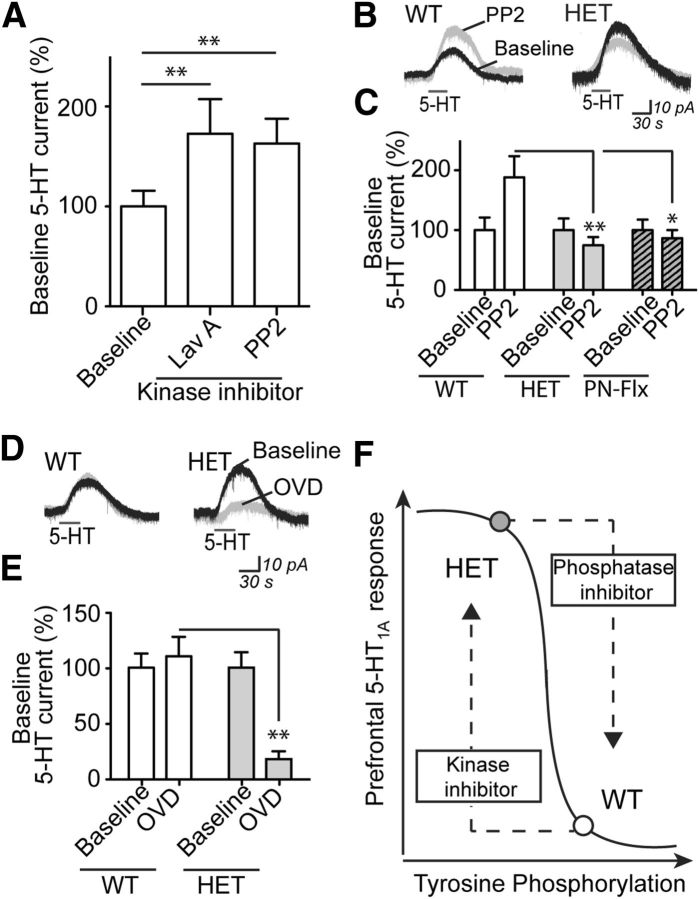
Figure 3.
Inhibition of 5-HT1A electrophysiological responses by tyrosine kinase activity is inefficient, but not lost, in mice with 5-HTT disruption. A, The 5-HT1A responses from WT neurons in the absence (combined baseline, n = 51) or presence of either tyrosine kinase inhibitors lavendustin A (10 μm, Lav A, n = 19) or PP2 (10 μm, n = 41). Averaged recordings (B) and quantification (C) of the 5-HT1A response in the absence or presence of Src family tyrosine kinase inhibitor PP2 in neurons from WT (baseline, n = 22; PP2, n = 17), HET (baseline, n = 18; PP2, n = 18), and PN-FLX (baseline, n = 13; PP2, n = 19) mice. In graph, baseline-normalized data are shown to demonstrate that the effect of PP2 differs significantly across groups. Independent replications of group differences in 5-HT response: WT versus HET or PN-FLX (p < 0.01). Averaged recordings (D) and quantification (E) of the 5-HT1A responses in the absence or presence of tyrosine phosphatase inhibitor OVD (1 mm) from neurons of littermate WT (baseline, n = 25; OVD, n = 20) and HET (baseline, n = 13; PP2, n = 14) mice. In graph, baseline-normalized data are shown to demonstrate that the effect of OVD differs significantly between groups. Independent replication of group difference in 5-HT response: WT versus HET (p = 0.0001). F, Schematic illustrating the working model of the relationship between prefrontal 5-HT1A responses and tyrosine phosphorylation in 5-HTT WT and HET mice. *p < 0.05. **p < 0.01.

